References

Zuchelli G.Italy: Quintessence; 2019
Chu SJ, Tan JH, Stappert CF Gingival zenith positions and levels of the maxillary anterior dentition. J Esthet Restor Dent. 2009; 21:113-120 https://doi.org/10.1111/j.1708-8240.2009.00242.x
Chu SJ. Range and mean distribution frequency of individual tooth width of the maxillary anterior dentition. Pract Proced Aesthet Dent. 2007; 19:209-215
Cohen ES.Hamilton, ON, Canada: BC Decker Inc; 2007
Caton J, Armitage G, Berglundh T A new classification scheme for periodontal and peri-implant diseases and conditions – Introduction and key changes from the 1999 classification. J Clin Periodontol. 2018; 45:S1-S8 https://doi.org/10.1111/jcpe.12935
Garguilo AW, Wentz FM, Orban B. Dimensions and relations of the dentogingival junction in humans. J Periodontol. 1961; 32 https://doi.org/10.1902/JOP.1961.32.3.261
Goldman HM, Cohen DW.St Louis, MO, USA: CV Mosby; 1968
Coslet GJ, Vanarsdall R, Weisgold A. Diagnosis and classification of delayed passive eruption of the dentogingival junction in the adult. Alpha Omegan. 1977; 10:24-28
Nart J, Carrio N, Valles C Prevalence of altered passive eruption in orthodontically treated and untreated patients. J Periodontol. 2014; 85:E348-53 https://doi.org/10.1902/jop.2014.140264
Wise MD. Stability of gingival crest after gingival surgery and before anterior crown placement. J Prosthet Dent. 1985; 53:20-23 https://doi.org/10.1016/0022-3913(85)90057-5

Aesthetic crown lengthening

From Volume 1, Issue 1, March 2024 | Pages 27-32

Authors

Reena Wadia

BDS Hons (Lond), MJDF RCS (Eng), MClinDent (Perio), MPerio RCS (Edin), FHEA

Founder and Principal Specialist Periodontist, RW Perio and Perio School, 75 Harley Street, London

Articles by Reena Wadia

Email Reena Wadia

Abstract

Ideal smile aesthetics includes ‘pink’ aesthetics and aesthetic crown lengthening surgery is an integral surgical procedure that is commonly used to achieve optimal overall aesthetics. Understanding the reasons for a ‘gummy’ smile, or gingival excess, is key. A thorough assessment and careful treatment planning are required for successful results. Crown lengthening can be carried out as a stand-alone procedure or as part of a wider restorative treatment plan.

CPD/Clinical Relevance: This article discusses the ideal ‘pink’ aesthetics, indications for crown lengthening as well as the treatment sequence.

Article

With an increase in patient demand for achieving the most optimal smile aesthetics, the field of periodontology has also evolved to have a greater focus on periodontal plastic surgical procedures to achieve the required ‘pink’ aesthetics as part of this. One of the most popular treatments is aesthetic crown lengthening. Crown lengthening surgery can be used to address ‘gummy smiles’, uneven gingival contours and can enhance the appearance of restorations placed within the aesthetic zone.

Ideal ‘pink’ aesthetics

The smile

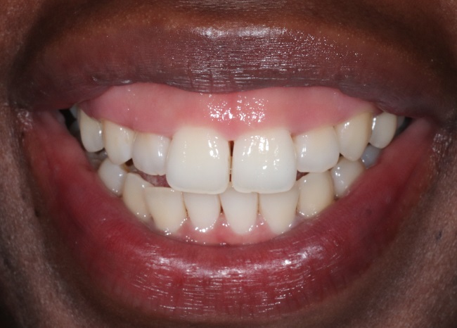
A smile is generally defined as pleasant when it exposes the entirety of the maxillary teeth along with approximately 1 mm of facial gingiva. Gingival exposure of up to 2–3 mm is normally found acceptable, whereas patients are usually dissatisfied with any greater exposure (>3 mm).1 Patients with a high smile line who expose a large band of gingiva may be classified as having a ‘gummy’ smile (Figure 1).

Figure 1.

Patient with what could be considered a ‘gummy’ smile.

The gingivae

The ideal gingival architecture consists of several features:

  • Gingival zenith position: The gingival zenith is the most apical point of the gingival outline. According to Chu et al, the gingival zeniths of the maxillary central incisors should be approximately 1 mm distal to the midline of the crown, the zeniths of the lateral incisors 0.4 mm distal, and the zeniths of the canine usually centralised along the long axis (Figure 2).2
  • Relative gingival margins: Apico-coronally, the zeniths of the lateral incisors should be 1 mm coronal to central incisors and canines (Figure 2).
Figure 2.

Diagram demonstrating the ideal gingival architecture.

The gingival aesthetic line is defined as the line joining the tangents of the central incisor and canine zeniths. The gingival aesthetic line angle is that formed at the intersection of this line to the maxillary dental midline. The ideal angle is between 45° and 90° (Figure 3). In the same respect, gingival symmetry between the right and left side of the mouth is an important consideration.

Figure 3.

Diagram demonstrating the ideal gingival aesthetic line angle.

One should also consider the ideal height-to-width tooth ratios: the central incisor is 80%, the lateral incisor is 70% and the canine is 75%.3

Crown lengthening: definitions and indications

Crown lengthening has been described as the surgical removal of hard and soft periodontal tissue to gain supragingival tooth length, allowing for longer clinical crowns and the re-establishment of the biological width.4 Patients now frequently refer to this procedure as a ‘gum lift’. The term biological width has now been replaced with the term supracrestal tissue attachment in the updated periodontal classification.5 This encompasses the junctional epithelium and supracrestal connective tissue (Figure 4). This should be approximately 2 mm.6 If the supracrestal tissue attachment is not respected or recreated, this will result in the unwanted consequences of rebound of the gingival tissue, gingival inflammation, pocketing recession and/or bone loss.

Figure 4.

Diagram demonstrating supracrestal tissue attachment, encompassing the junctional epithelium and supracrestal connective tissue.

Crown lengthening can be categorised as aesthetic or restorative/functional. Aesthetic crown lengthening focuses on correcting excessive gingival show, short clinical crown heights and uneven gingival contours. Restorative/functional crown lengthening is required when there is inadequate tooth structure for restorative treatment, to access subgingival fractures/caries/perforations or to relocate crown margins impinging on the supracrestal tissue attachment.

Gingival excess

Before prescribing aesthetic crown lengthening, a diagnosis is required. According to the most recent classification scheme, under the umbrella of mucogingival deformities and conditions, the term gingival excess has been used.5 This may form part of the diagnosis and is associated with several causes. These may involve different anatomical structures, such as the teeth/periodontium, lips, jaw/facial structures, either individually or in combination. The most common causes include altered passive eruption, dento-alveolar extrusion following attritive tooth wear, vertical maxillary excess and a short or hypermobile upper lip. It is important to establish the exact cause(s) as not all will be well suited to aesthetic crown lengthening surgical procedures. For example, conditions such as vertical maxillary excess may require maxillofacial or orthognathic surgery to resolve fully, and a hypermobile lip may benefit from botulinum toxin injections or lip repositioning surgery. In this article, we focus on altered passive eruption.

Altered passive eruption is one of the most common conditions that can be addressed with aesthetic crown lengthening. Passive eruption commences after the tooth's anatomical crown has fully erupted and is characterised by the apical shift of the dentogingival junction. The length of the clinical crown increases as the epithelial attachment migrates apically. Apical migration of the dentogingival junction continues until it reaches a physiological distance of 0.5–2.0 mm coronal to the cemento-enamel junction (CEJ). If alterations occur during the passive phase of tooth eruption, the gingival margin fails to retract to the full extent, giving rise to the phenomenon of altered or delayed passive eruption. Altered passive eruption was first defined by Goldman and Cohen.7 Later, a classification system, based on the relationship between the gingivae and the underlying alveolar bone, was developed (Table 1).8 This condition is not uncommon, and altered passive eruption may affect approximately 35% of the population.9 The gingival margin position in relation to the CEJ and buccal bone crest, as well as the crown–root–alveolar crest relationships, needs to be evaluated to ascertain whether the gingival excess, or ‘gummy’ smile, is due to altered passive eruption.

Table 1.

Classification of altered passive eruption.

Classification Description Wide band of keratinised tissue (>2 mm) Distance between CEJ and alveolar bone crest 1.5–2 mm Wide band of keratinised tissue (>2 mm) CEJ and alveolar bone crest close or at the same level Narrow band of keratinised tissue (≤2 mm) Distance between CEJ and alveolar bone crest 1.5–2 mm Narrow band of keratinised tissue (≤2 mm) CEJ and alveolar bone crest close or at the same level

The treatment sequence

The planning

The planning phase for aesthetic crown lengthening is crucial for a successful long-term outcome and meeting patient expectations. First, it is important to ensure, or attain, periodontal health and optimal plaque control prior to this elective surgical procedure. Therefore, if after a thorough periodontal examination, the patient has been diagnosed with gingivitis or periodontitis, this should be treated first (Figure 5). Other local factors, such as the crown:root ratio, level of the furcation entrance, root proximity and endodontic status and restorability of the tooth should also be assessed. If any endodontic treatment is required, this should be completed prior to the crown lengthening.

a

b

c

Figure 5.

Before and after photographs of a patient treated for gingivitis prior to crown lengthening. (a) Pre-treatment; (b) following non-surgical periodontal therapy; and (c) following crown lengthening surgery.

The next step is to determine the reference point or finish line. This is usually the CEJ for altered passive eruption cases, but could also be a reference point on the tooth, existing/provisional restorations or that guided by a surgical stent. Understanding the ideal ‘pink’ aesthetics and then evaluating the current architecture and extent of the smile will determine how much and how many teeth will require crown lengthening. Looking at the face as a whole and the interpupillary line for facial symmetry can be helpful. The extent of crown lengthening may also be dictated by the restorative treatment plan. With aesthetic crown lengthening cases, it is important to involve the patient to ensure expectations are met or corrected to be more realistic prior to completing the surgery. Visualisation of the end result through annotated photographs (Figure 6), composite mock-ups in the mouth and wax-ups with stents can all be helpful (Figure 7). Digital smile design will form a part of this process (Figure 8). Chu probes are available to help assess ideal proportions of the teeth (Figure 9). These will all also be helpful for the clinician when it comes to completing the surgical procedure. Cone-beam compute tomographs (CBCTs), if available for other reasons, can be a useful diagnostic and planning tool, but are currently not commonly completed for the sole purpose of treatment planning for crown lengthening.

a

b

c

c

Figure 6.

Before and after photographs of a patient undergoing crown lengthing. (a) Pre-treatment; (b) pre-treatment using annotations for visualisation of the end result; and (c) post-treatment photograph.

a

b

c

d

Figure 7.

Composite mock-ups in the mouth and wax-ups with stents can all be helpful in cases of aesthetic crown lengthening. (a) Digital pre-operative model; (b) digital diagnostic wax-up; (c) study casts; and (d) surgical guide or stent. Courtesy of Dr Anish Berry, the referring dentist.

Figure 8.

Digital smile design will form a part of the treatment sequence for aesthetic crown lengthening. Courtesy of Dr Arman Barfeie, the referring dentist.

Figure 9.

Chu probes can be useful for assessing the ideal proportions of the teeth.

The amount of keratinised tissue needs to be assessed because this will determine the surgical approach: resective versus apically positioning tissues (or a combination of both). Ideally, it is important to leave at least 2 mm of keratinised tissue post-surgery to enable the patient to maintain an optimal level of plaque control more comfortably than if only lining mucosa remained.

The position of the bone and the distance between the bone level and reference point will then determine the amount of bone removal that will be required (if any), respecting the supracrestal tissue attachment. The supracrestal tissue attachment should be approximately 2 mm, and if the gingival sulcus depth is added in, the aim is to have arpproximately 3 mm between the reference point (final restoration margin/gingival margin) and bone level. If that already exists, then no bone removal is required and gingivectomy only will be performed. If it does not, then bone removal is necessary to avoid encroachment into the supracrestal tissue attachment and its associated negative consequences. Good quality periapical radiographs and bone sounding will be useful to help determine the position of the bone and the supracrestal tissue height. The use of GP points and radiographs may be a useful adjunctive technique to determine the clinical and anatomical crown height in comparison to the bone level (Figure 10). On most occasions, the exact position of the bone will be reconfirmed when a flap is raised at the time of the surgical procedure unless one is sure that no bone removal is required.

Figure 10.

The use of GP points and radiographs may be a useful adjunctive technique to determine the clinical and anatomical crown height in comparison to the bone level. Courtesy of Dr Devan Raindi.

Table 2 summarises the various surgical approaches.

Table 2.

Summary of the various surgical approaches.

The surgery

In aesthetic crown lengthening, generally only a buccal flap is raised and only buccal bone removal is necessary. However, with restorative/functional crown lengthening, both a buccal and palatal/lingual flap is raised because circumferential bone removal is usually necessary for all surfaces. Initially, bleeding points are completed (Figure 11) at the new gingival margin position, followed by a superficial scoring incision in the epithelium and only once happy with this incision is a full thickness incision completed. To reduce the chances of ‘black triangles’ developing, the interproximal tissue can be thinned rather than fully reflecting or preserved by not including it in the flap design. Vertical incisions tend to be avoided to minimise any scarring. A 15c blade can be used for the incisions, or a laser if preferred.

Figure 11.

Initial bleeding points are completed.

If bone removal, and specifically ostectomy, is required, the aim is to remove sufficient bone to re-establish the supracrestal tissue attachment at its new position.

It is also key to ensure there is a gradual rise and fall in the profile of the osseous crest to recreate a positive architecture so osteoplasty may also be required.

If there are any bony exostoses that are visible on smiling these may also need to be addressed through osteoplasty (Figure 12). Bone removal can be completed via suitable anti-retraction micromotor or piezoelectric handpieces/burs and may be supplemented with hand chisels/curettes. It is important to use the correct equipment to prevent the risk of surgical emphysema.

Figure 12. 

Bony exostoses that are visible on smiling may also need to be addressed through osteoplasty.

The suture technique is usually single interrupted or mattress sutures. Usually, if the gingival tissue needs to be everted in aesthetic cases (to avoid ‘black triangles’) then an internal mattress suture is ideal (Figure 13). If the papilla is wide or there is a diastema then a horizontal internal mattress suture would be most suitable and if narrow, then a vertical internal mattress suture. Thin sutures such as a 5-0 or 6-0 will allow for the best healing. The exact material is operator preference. Unlike restorative/functional crown lengthening, periodontal dressings are not used in aesthetic crown lengthening cases. The sutures are removed approximately 2 weeks following the surgical procedure. Regular analgesia are advised as needed, along with a soft diet for a 4–5 days and use of an antiseptic mouthrinse (avoiding brushing the surgical site) for 2 weeks. In altered passive eruption cases, only the crown of the tooth is usually exposed. Sensitivity is usually not an issue, so no further action is usually required in this 2-week period. Often in other cases, especially restorative cases, some root exposure may be present and any resultant sensitivity may need to be addressed as required.

a

b

c

Figure 13.

An internal mattress suture is ideal if the gingival tissue needs to be everted in aesthetic cases. (a) Pre-operative photograph; (b) immediate post-operative internal mattress sutures using 6-0 vicryl; and (c) photograph taken 1 month post-operatively.

Further restorative treatment

Altered passive eruption cases may not always require restorative work following surgery unless there is an additional need to improve the tooth aesthetics (Figures 1419). If crown lengthening has been completed for other restorative cases, such as tooth wear cases following dento-alveolar extrusion, then a restorative phase is likely to follow. Provisional restorations can be placed after suture removal, but for those teeth in the aesthetic zone, there is limited evidence to suggest that it may be wise to wait 6 months prior to the placement of the definitive restorations because the position of the gingival crest may continue to change until then (Figure 20).10 For this reason, lab-based provisional restorations are commonly used in this interim period. Coronal displacement of the gingival margin appears to be more pronounced in thick gingival phenotypes, but all patients should be consented for a revision surgery in case it is needed. Usually if this is needed and the bone level is at the correct level, it may be that a gingivectomy is sufficient.

a

b

Figure 14.

Crown lengthening surgery for the UR5–UL5. (a) Pre-operative; (b) post-operative.

a


b

Figure 15.

Crown lengthening surgery for the UR5–UL5. (a) Pre-operative; (b) post-operative.

a

b

Figure 16.

Crown lengthening surgery for the UR5–UL5. (a) Pre-operative; (b) post-operative.

a

b

Figure 17.

Crown lengthening surgery for the UR5–UL5. (a) Pre-crown lengthening, post-orthodontics; (b) post-crown lengthening.

a

b

Figure 18.

Crown lengthening surgery for the UR5–UL5. (a) Pre-crown lengthening; (b) post-crown lengthening, but pre-whitening and restorative treatment.

a

b

Figure 19.

Crown lengthening surgery for the UR5–UL5. (a) Pre-treatment; (b) post-treatment. Crown lengthening surgery by Dr Reena Wadia, restorative treatment by Dr Sahil Patel.

a

b

Figure 20.

(a) Pre-treatment;


(b) post treatment. Crown lengthening and recession surgery by Dr Reena Wadia, restorative work by Dr Sahil Patel.


Conclusions

Assessment of the ‘pink’ aesthetics is becoming just as important as the ‘white’/tooth aesthetics to ensure the most optimal aesthetic outcomes for our patients. Aesthetic crown lengthening can have a significant impact on overall smile aesthetics and impact an individual's confidence. It can be used as a stand-alone treatment or part of multidisciplinary cases, such as post-orthodontics or as part of a combined restorative treatment plan. Understanding the biological concepts, accurate treatment planning and careful execution are critical for successful results.